What are age spots, and can they be safely removed?
Age spots are flat, benign areas of increased skin pigmentation that usually develop after years of sun exposure. In many cases, they do not need treatment for medical reasons, but a proper skin assessment matters because some pigmented marks can look similar to more concerning lesions. Once the diagnosis is clear, options such as prescription creams, cryotherapy, and certain laser treatments may reduce or remove them, depending on the mark itself and the person’s skin type.
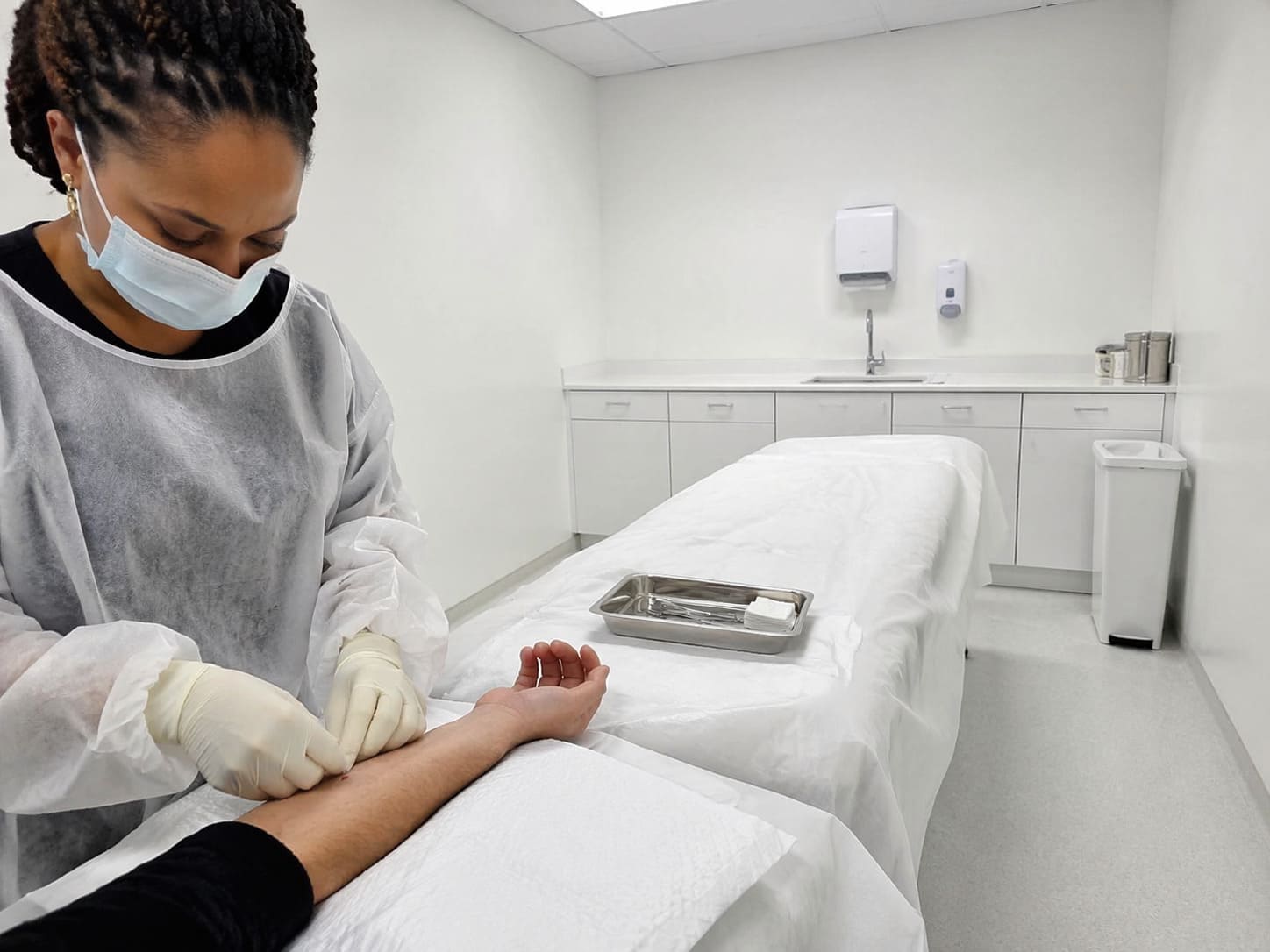
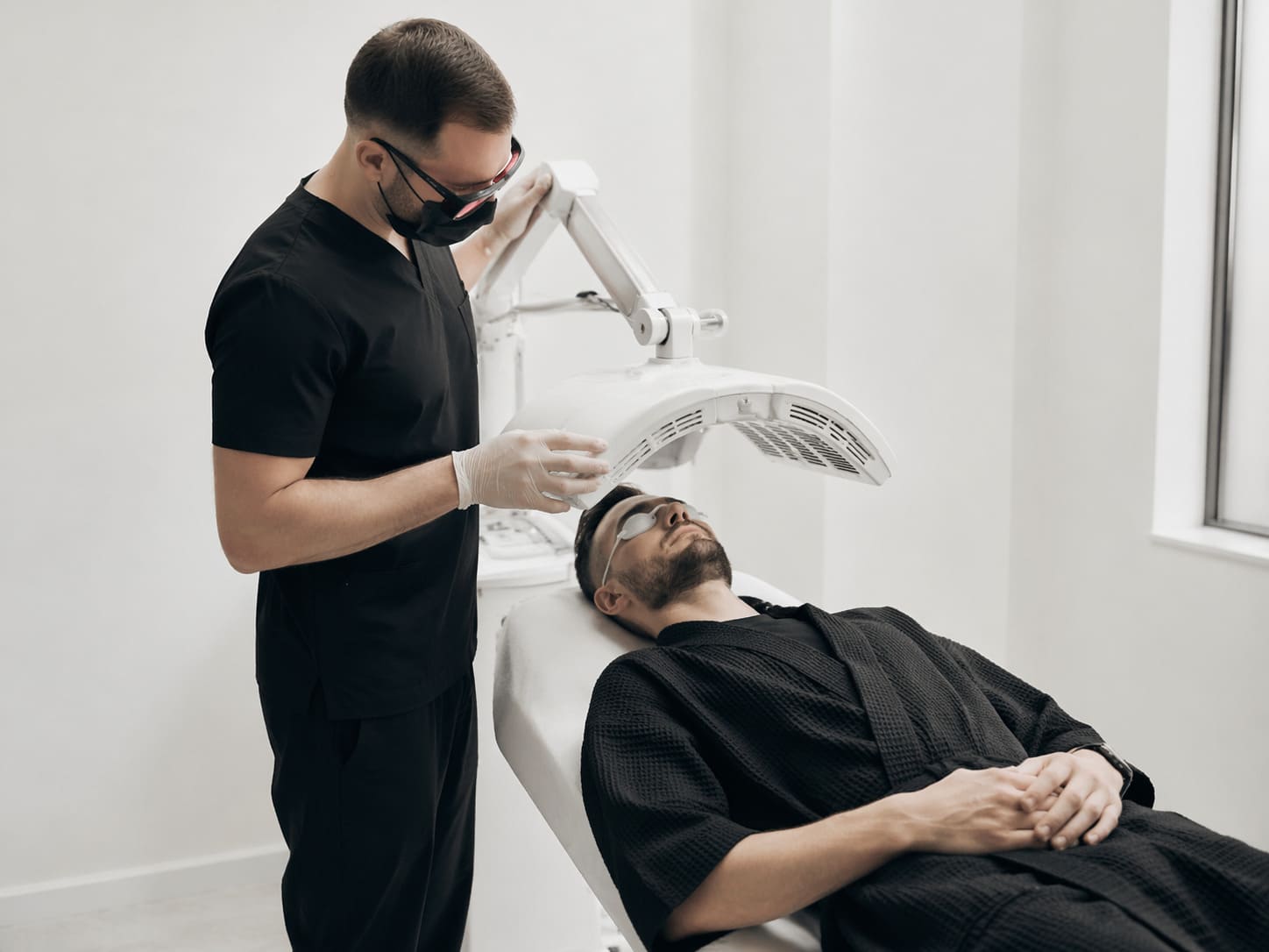
Skin Lesion Removal Procedure (3) – Illustrative Image
What Is In This Article
Understanding Age Spots: What They Are and Why They Matter
Age spots, which are also called liver spots or solar lentigines, are common brown spots that tend to appear on skin that has had repeated ultraviolet exposure. Hands, face, shoulders, chest, and forearms are typical sites. The colour comes from localised increases in melanin, which is the pigment that gives skin its natural tone.
Despite the name, these marks are not related to the liver. They are usually part of age-related skin changes linked to cumulative sun damage and skin ageing. NHS information and guidance from the British Association of Dermatologists both support the broader point that sun exposure plays a central role in many changes in skin pigmentation.
Confusion often arises because age spots are only one type of pigmented lesion. A flat brown patch may be harmless, but another mark with a similar colour may need closer assessment.
- Age spots are usually flat, clearly defined, and uniform in colour.
- Freckles often darken with sun exposure and may fade when sun exposure drops.
- Moles can be raised or flat and may have a different structure from solar lentigines.
- Some skin cancers, including melanoma, may begin as new or changing pigmented lesions.
Visible skin changes matter for two reasons. First, any change in colour, shape, or pattern can raise a valid question about skin cancer risk. Second, marks on the face or hands are hard to ignore in daily life, particularly for people in public-facing or professional roles. That practical impact often explains why people seek advice even when the lesion itself is benign.
The Main Causes of Age Spots
Age spots develop mainly because skin stores the effects of sun exposure over time. A single bright day does not create them on its own, but years of ultraviolet light can alter how pigment is produced and distributed.
Cumulative UV exposure
Ultraviolet light acts as the main trigger for melanin production. Melanin helps protect the skin, but repeated exposure can lead to patches where pigment becomes concentrated. That process is one reason solar lentigines appear more often on areas that receive regular daylight, including during commuting, outdoor exercise, or seated time near windows.
City living does not remove that risk. Walking between stations, eating lunch outdoors, cycling to work, and weekend time in stronger seasonal sun all add to cumulative exposure. Photoageing often reflects the sum of these routine patterns, not a single obvious event.
Skin ageing and cell turnover
As skin ages, repair processes and skin cell turnover change. Pigment can become less evenly distributed, which means that localised brown patches become more likely. That is why age spots are more common in older adults, even though the underlying sun exposure usually began much earlier.
Skin type and genetics
Individual skin behaviour also matters. People with lighter skin types often develop visible sun-related pigmentation more easily, although age spots can occur across different skin tones. The Fitzpatrick skin type classification is sometimes used in dermatology to assess how skin responds to sun exposure and to guide treatment suitability.
Family patterns may also influence how readily someone develops pigmentation changes. Genetics does not remove the role of ultraviolet exposure, but it can affect who develops noticeable marks sooner.
Common risk factors
- Long-term sun exposure without consistent sun protection
- Outdoor work or outdoor hobbies
- Fairer skin that burns easily
- Increasing age
- A history of frequent tanning or sunburn
None of this means age spots are unavoidable. It does mean that the causes of age spots are usually cumulative, which is why prevention has to be routine rather than occasional.

Mole Check Assessment – Illustrative Image
How Age Spots Are Diagnosed and Assessed Clinically
A new brown mark on the hand may look harmless in the mirror, yet uncertainty often remains if it has changed, become darker, or appeared later in life. That is where skin lesion assessment becomes useful. The aim is not simply to label a spot, but to distinguish a benign lesion from one that needs further investigation.
During a dermatology consultation, the clinician usually begins with a history. They may ask when the mark appeared, whether it has changed, whether it itches or bleeds, and whether there is any personal or family history of skin cancer. After that, the lesion is examined directly.
A structured assessment often includes:
- Visual inspection of the lesion and the surrounding skin.
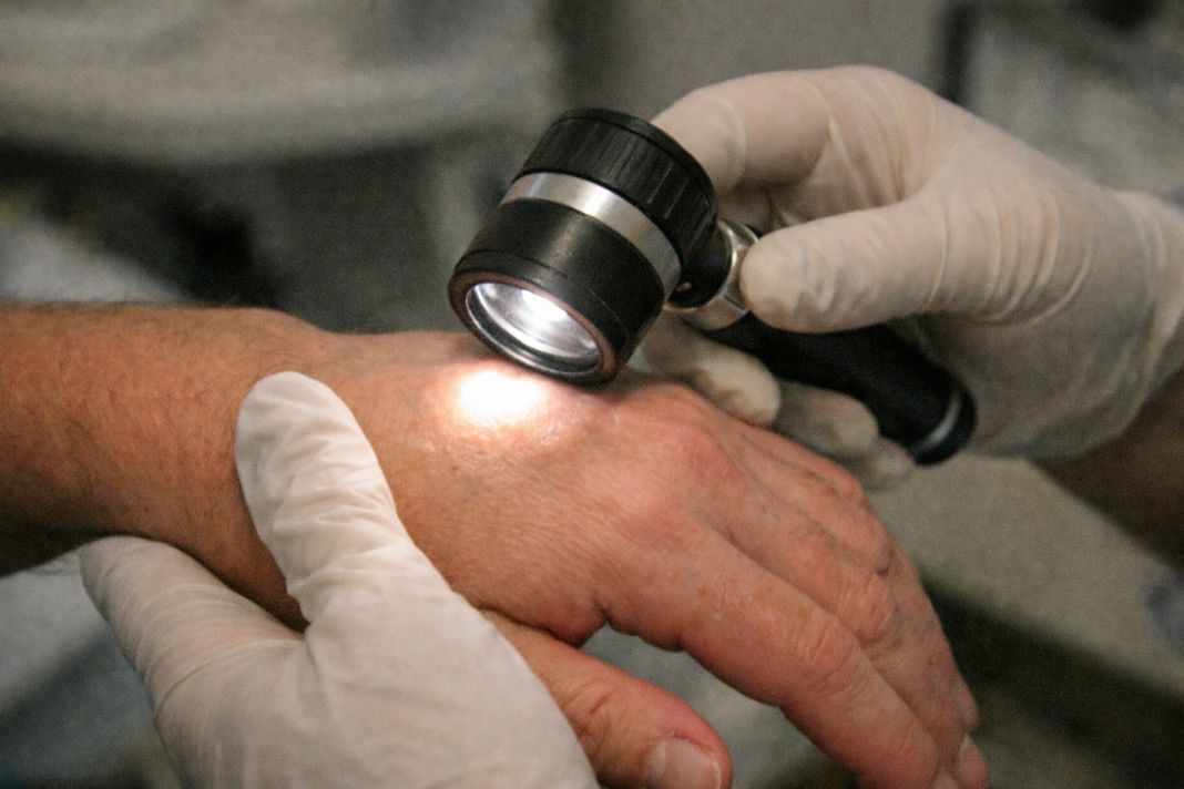
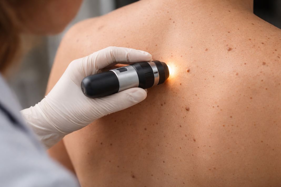
- Dermatoscopy, which uses a handheld device to examine pigment patterns more closely.
- Review of whether the lesion has features that fit a benign diagnosis such as solar lentigo.
- Consideration of biopsy or removal if the appearance is unclear or concerning.
Dermatoscopy is especially useful because pigment patterns that look similar to the naked eye can behave very differently under magnification. Consultant dermatologists use it to assess colour distribution, borders, and internal structures that help separate benign from malignant lesions.
Self-monitoring still has a place, but it has limits. A flat brown patch that stays stable over time may simply need observation. A mark that changes in shape, colour, size, or sensation should be reviewed professionally instead of being assumed to be an age spot. In a clinic setting such as Future Care Medical, that assessment can sit within a wider skin screening pathway if other lesions also need review.
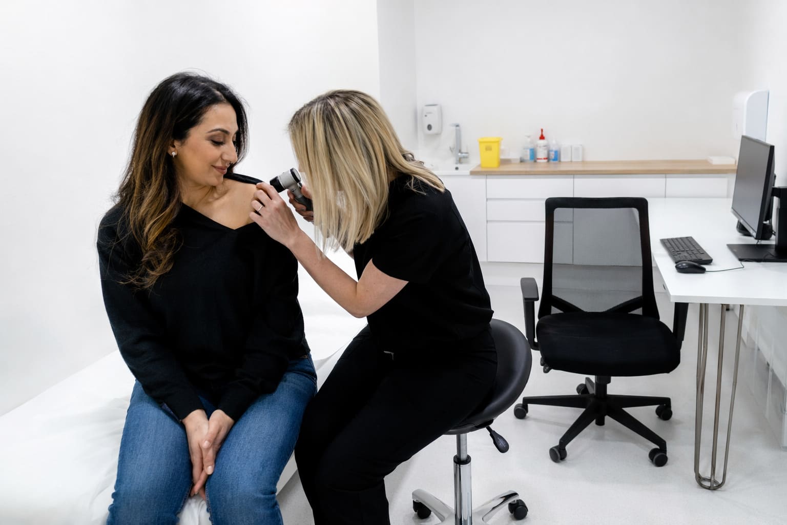
Collarbone Mole Check – Illustrative Image
What Actually Removes Age Spots? Evidence-Based Treatment Options
Removal depends on one prior step: confirming that the lesion is actually an age spot. Once that diagnosis is established, treatment can focus on pigment reduction or lesion destruction, depending on the size, site, skin type, and clinical judgement.
Some options lighten the excess pigment gradually. Others physically remove or destroy the pigmented tissue. No approach suits every patient or every lesion, and realistic expectations matter because response can vary.
Prescription creams
Certain prescription treatments may reduce pigmentation over time. These can include agents used to alter pigment production or speed skin cell turnover, such as hydroquinone or retinoids where clinically appropriate. They are generally more relevant for selected superficial pigmentation and usually require consistent use over weeks or months.
Topical treatment is not a quick fix. Irritation, uneven lightening, and limited response can occur, particularly if the diagnosis is wrong or sun protection is poor during treatment.
Laser and light-based treatments
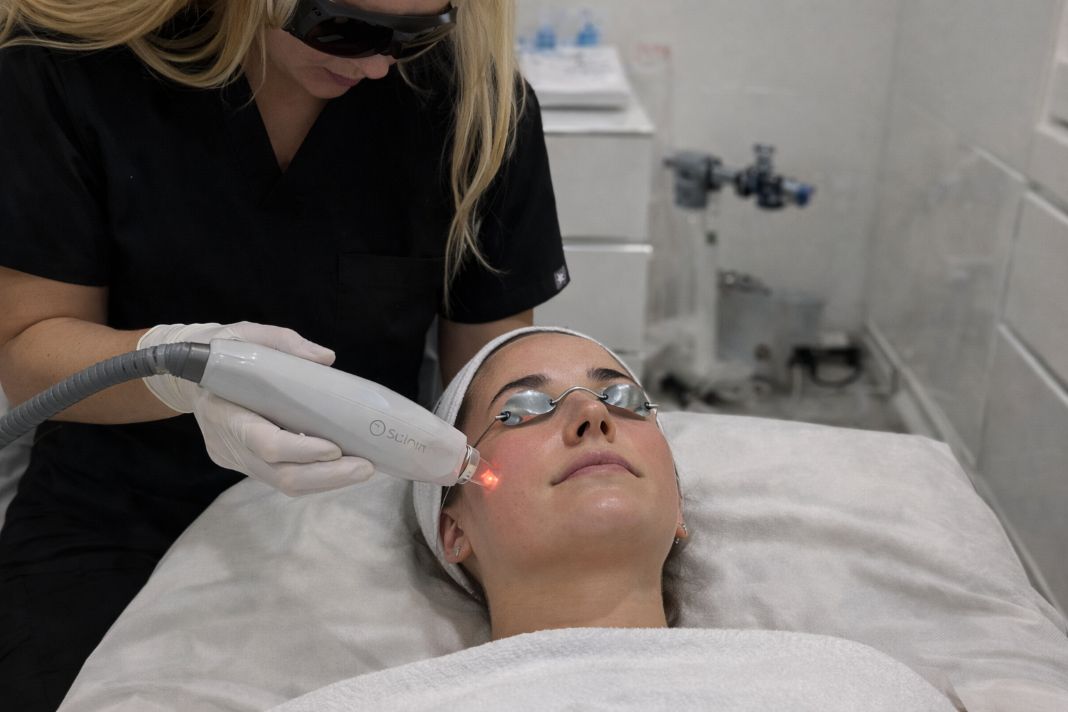
Laser for age spots can be effective in selected cases because targeted energy is absorbed by pigment within the skin. The body then clears some of the disrupted pigment over time. Suitability depends on the exact lesion, skin tone, and the risk of post-inflammatory pigmentation.
Where a clinic already offers laser-based skin treatments, the lesion still needs medical assessment before any pigment treatment is chosen. At Future Care Medical, doctor-led skin treatment pathways and dermatology input help determine whether a brown spot is appropriate for treatment at all. If a patient is being assessed for wider skin rejuvenation concerns, relevant laser pricing within the clinic includes PicoSure Pro at £480.00, although a pigmented lesion still requires diagnosis before any treatment plan is matched to it.
Cryotherapy
Cryotherapy uses controlled freezing to destroy superficial abnormal tissue. It is sometimes used for benign pigmented lesions, including certain age spots. The treated area then heals over time, although temporary crusting, colour change, or incomplete clearance can happen.
Hands and forearms are common sites where cryotherapy may be considered, but the cosmetic result is not always predictable. For that reason, lesion type and skin tone both influence whether it is a sensible option.
Minor procedures and biopsy
If a lesion is atypical, raised, changing, or diagnostically uncertain, removal may be carried out as a minor procedure with tissue sent for analysis. That is not standard treatment for every age spot, but it becomes relevant when benign and malignant appearances overlap.
The most important point is simple. Effective age spot treatment begins with accurate diagnosis, because the right treatment for a solar lentigo is very different from the right management for a suspicious pigmented lesion.
Prevention and Long-Term Skin Health
Daily sun protection is the main practical step for preventing age spots and reducing further pigment change. Even in Central London, cumulative daylight exposure adds up through commuting, walking between offices, or sitting near windows for long periods.
A workable routine usually includes the following:
- Use broad-spectrum sunscreen on exposed skin, particularly face, hands, chest, and forearms.
- Reapply if you are outdoors for extended periods.
- Wear hats or protective clothing during stronger sun exposure.
- Pay attention to new or changing pigmented marks.
- Arrange clinical review if a lesion looks different from your usual pattern.
Public health campaigns, NHS advice, and recommendations from the British Association of Dermatologists all support the broader principle that sun protection is a long-term behaviour rather than a holiday-only measure.
Regular skin monitoring also matters. Many people know their moles and brown spots well enough to notice change, which means that new asymmetry, multiple colours, crusting, bleeding, or persistent itch should stand out. Early review is useful because pigmented lesions are easier to assess when change is recognised clearly and not months later from memory alone.

Skin Lesion Removal Procedure – Illustrative Image
Common Misconceptions and What to Remember
Several age spot myths persist, and some of them lead people either to ignore a concerning lesion or to spend time on treatments that are unlikely to help.
An age spot is not the same as skin cancer, but it is also not wise to assume that every brown spot is harmless. Appearance alone can be misleading, which is why consultant dermatologists rely on clinical examination and dermatoscopy instead of guesswork.
Over-the-counter products may promise to remove brown spots, yet unproven remedies can irritate the skin without addressing the lesion properly. A cream cannot safely replace clinical evaluation if the diagnosis is uncertain.
Age spots are a normal feature of skin ageing for many adults, especially on sun-exposed areas. That normality does not mean every lesion should be ignored. The sensible approach is to view them in context: stable, benign marks may be monitored or treated for pigment reduction, whereas anything new, changing, or unusual deserves proper assessment first.